Click to suport!

Friday, October 18, 2013

Biomaterial processes: DIY tissue engineering for dummies

Checking on the growth of my new cell cultures in the Pelling Lab.
I always vowed to myself throughout my academic career that I would never, ever, ever do a PhD. It seemed ludicrous. It seemed like way too much academia, and I don't exactly love the structure of academia. I didn't think I would ever have a single idea that could sustain 5-7 years worth of research. Plus, an MFA is officially a terminal degree for artists. However, the tissue engineering workshop at the Pelling Lab changed my thinking completely. I gained a very clear sense that my entire art practice thus far has led me to this work, and that everything I've ever done now makes perfect sense. Then I discovered that SymbioticA has a PhD program in creative tissue engineering. Oh, dear.

Oron Catts is one of the world leaders and innovators of the intersection between tissue engineering and art. I really enjoyed working with him at the Pelling Lab. It wasn't just the technical processes he so openly and generously shared, but also his philosophy towards science and scientific research: that the ongoing mystification and media hype needs to be debunked, and that scientific research is accessible, feasible and important for artists. Also, artists are important for science. He did, however, inform us that we were quite privileged in being selected to participate in the workshop and in actually being given this kind of access so openly.

One friend, with an academic background in sciences, asked me outright how it was that I, someone with no training whatsoever in science, had access to these kinds of labs (type II containment labs necessary for working with biohazardous materials), and this kind of highly specialized work. I explained that through this workshop, I was paired up with biologists to learn the 'craft' (as Oron calls it) of tissue engineering. For me, as an artist with a background in fine craft practices, this is speaking my language. Interdisciplinary practice is hot in the art world and in the research funding world so more and more we are seeing different fields of research being paired up with art practices. This is ultimately very enriching. Oron is interested in disseminating information that will enable artists to use semi-living matter (definition: that which comes from a living entity but can only exist in a laboratory environment) as art material. This art material is essentially disembodied life. And, as an artist, I can muck about with wet biology processes to dream up and create a new body for that disembodied life! I can hear that [here unnamed] church congregation praying once again for the salvation of my soul. I joke about that but in all seriousness, there are ethical implications with this type of work, and those problematics are extremely interesting to me, and part of the work that Oron explored with us. More on that later.

In the first day of the workshop, we learned to construct DIY incubators to grow biological cultures in, and air purifier hoods for creating a sterile work environment. Oron brought everyone to the labs, showed us a pile of materials that he'd selected for us, and instructed us to collaboratively figure out how to use the materials to build the lab equipment we'd need for growing cell cultures. 

The inside of our DIY incubator.
I was in the incubator working group. We riffled through the pile and chose a styrofoam cooler, a heating pad, a heating wire with a temperature sensor, a thermostat, a digital thermometer/ barometer, and foil tape. We also had scissors and a hand saw. None of us had any experience with these things previous to this. With those materials and basic pieces of equipment, we turned the cooler into a successfully functioning incubator that kept a fairly evenly-regulated temperature of 38˚ (body temperature). Animal cells need to be kept at body temperature to stay alive and grow. Our incubator had a plastic window in the door, built from the packaging that the thermostat came in. Oron was tough and gave us a hypothetical grade of 51%. Our incubator was not cell culture quality, he said, but we could grow mushrooms or culture yogurt/bacteria in it if we really wanted to. In truth, the materials he gave us would likely never produce an incubator suitable for cell cultures, but the exercise of collaborating to put one together with no previous knowledge whatsoever was empowering.

In the second day, we learned the processes of decellularization and creating new cell cultures. Decellularization is absolutely fascinating. Animal tissues, when all cells are removed, exist as nothing but a scaffolding of collagen strands. Plant matter (as opposed to animal tissue), when decellularized, becomes cellulose scaffolding. Animal = collagen. Plant = cellulose. Those are the matrices that all living matter grow on, the underlying structures that hold everything together. I thought of Marvin the Martian in Bugs Bunny cartoons, with his disintegrator ray gun, and imagined that it must simply eliminate collagen. Without the collagen scaffolding, we might collapse into a pile of loose cells with nowhere to go. 
A decellularized apple with only the cellulose scaffolding remaining.
A decellularized piece of tissue (pork) showing just the remaining collagen scaffolding (top) - it appears as a translucent, random mesh of fibres.
Decellularization is important because it allows you to create a raw material to work with, which can then be repopulated with any kind of living cell culture you want (FYI, many different types of living cells can be ordered online). One of the projects that was being worked on in the Pelling Lab was to repopulate an apple cellulose scaffolding with mouse muscle tissue. The apple was stripped of all its apple cells, then enculturated with the C2C12s (mouse muscle tissue cells), to produce an apple-shaped mouse muscle. They were interested in producing an apple that would twitch. Cells behave the way they're supposed to behave, meaning that heart cells will beat with the pulse of the heart they're taken from and muscle cells will know to expand and contract when stimulated. One thing that they discovered at the Pelling Lab was that a decellularized mouse heart repopulated with muscle tissue cells actually shrunk to half its size because the new muscle tissues contracted and pulled the heart form in. 

This is work in progress by Andrew Pelling's research crew. The container on the left is a decellularized apple and the container on the right is a decellularized apple that has been repopulated with C2C12s (mouse muscle tissue cells). It's in a red liquid because this is the nutrient solution necessary to feed and keep cell cultures alive and growing. The nutrient solution is made of fetal calf serum (blood) and antibiotics (since in vitro cell cultures do not have immune systems to protect them from contamination by diseases).
Once my piece of tissue was in the appropriate solution* in the magnetic stirrer to decellularize, I learned next how to create and grow my own cell cultures. We worked in the lab to create new HeLa (cervical cancer cells) and C2C12 cultures. HeLa cells are often used for testing in labs because they are so robust and grow very quickly. There is a history of complicated ethics around the use of HeLa cells (which were harvested from a dying woman without her knowledge or consent, or that of her family).

With a partner, I learned how to trypsonize an existing cell culture to separate the individual cells from each other (they grow in a tight, single layer on the bottom of the pitre dish), extract the free-floating cells from the old liquid, select a small sample of cells and transplant them to a new pitre dish, replenish the nutrient solution to feed them and establish a new culture (which has to go into the incubator to grow and thrive). This all has to be done in extremely sterile conditions in order to not contaminate the cells and cause them to sicken and die. Sick cell cultures will discolour the nutrient solution, so it is easy to tell which cultures are successful and which aren't. We came back to the lab the next day to check on our cultures - mine (a C2C12 culture) was growing remarkably well and looking very healthy, and I felt wondrously proud as I viewed them under the microscope. I'd grown my first in vitro life.

A successful C2C12 tissue culture grown collaboratively with my lab partner, Ottawa-based artist Daphne Enns, along with the cooperation of the cells themselves, of course. You cannot see the cells here because they are microscopic - all you can see is the hydrogel, but it is in fact swarming with hungry cells ready to proliferate under the right conditions.
You will notice that the above image of the cell culture indicates that it is suspended in hydrogel. Creating a hydrogel suspension was one of three techniques we learned for creating three-dimensional cell culture forms (decellularization/repopulating collagen scaffolding was the first technique). Hydrogel is extremely accessible, and in fact, Oron brought in a package of disposable baby diapers and showed us how to cut them open to collect the hydrogel beads that are embedded inside (used to cause baby urine to coagulate to keep the diapers 'dry'). So, the above cell culture is suspended in a hydrogel solution that I made from no-name diapers. Apropos for a culture of new (baby) cells, no? The laboratory (false) motherhood metaphor was not lost on me. As I mentioned, cell cultures grow in a single layer, typically, on the bottom of a pitre dish and stop reproducing when they run out of room. But, creating a hydrogel suspension and enculturating it with cells will provide the cells a 'shape' to grow into and through, and they will eventually take over the whole form in multiple layers. Were the cells pictured above allowed to continue to grow for an extended period of time, they would create a large, cylindrical muscle tissue the same shape as the entire pitre dish up to the edge of the hydrogel. This technique has fantastic possibilities. One could essentially create a mold of any shape and size, fill it with hydrogel suspension, enculturate it, incubate it and grow a semi-living form of one's own design.

The third technique for growing tissue in three dimensions is via a 3-D printer. In the Pelling Lab, there is a 3-D printer that extrudes a polymer cellulose in successive layers, building up whatever form you program it to print. We printed human vertebrae. Remember that cellulose is the plant-based matrix that cells can grow on, so this cellulose polymer is a friendly place for a new cell culture to grow, into the form you've printed. Eventually the polymer breaks down and the cells replace the underlying scaffold just as with the hydrogel.

These are the (two) vertebra we printed using the cellulose polymer extrusion process. If you look closely (or click the image to enlarge), you can see the layers of polymer that were built up. Polymer means 'plastic', but this is a vegetable- (cellulose-) based plastic that is biodegradable. Perfect for cells.
Some specialized labs are 3-D printing human organs. This is done with a different type of 3-D printer, one that prints cell-enculturated droplets of hydrogel into the form of a liver, etc. These experiments have been successful for reproducing organs in the lab.

A microscopic image of C2C12s on a computer screen at the Pelling Lab. These muscle tissue cells are being manipulated through a process of microscopic magnetic suction to attempt to align them all into the same direction or to change their shape. This is important for growing a muscle that will actually work properly to expand and contract in one direction.
Here an old iMac casing is being upcycled into an incubator (Pelling Lab).
This shows my (mostly) decellularized tissue after 24 hours in the magnetic stirrer, along with all of the others.
What happened with our living, thriving cell cultures? They were eventually disposed of (killed). We were asked first to think about what this really means and to collectively agree to allow those cultures to die. Personally, I did not have a problem with this decision, but ultimately, I did wistfully imagine taking it home and continuing to nurture this semi-living material the same as one would a plant or a pet or a child or any other living thing we care for, I suppose. This is impossible, of course, but I did still entertain the thought. Ideas of attachment to life and responsibility for that life, even in the process of engineering life in a false environment, is an important consideration, among others.

*I do have the recipe for the chemical bath that decellularizes tissue, along with all of the other recipes for feeding and sustaining cell cultures, etc.


Saturday, October 5, 2013

Gut Feeling

I've just ordered this box of catgut from eBay:


Catgut is a bit of a misnomer: it's actually a surgical thread made from sheep or goat intestines, not cat. Catgut eventually dissolves within the living body as tissue 'heals' and is therefore perfect for internal sutures. Here is how catgut used to be packaged and sold, historically (I'd love to have this object):

Click here to go to the medical antiques website.

I'll tell you why I've ordered this in a minute. Last week, I went to a local butcher and bought 10 meters of pig intestine (hog gut) for $10 and then knit it into a tight swatch. I had to knit it while it was still wet and malleable, meaning I knit it in a sink under a running faucet. Here's what it looked like afterwards:

You can see here that it is in the process of drying. The whole thing took about two days to dry.
Knitting intestine was relatively easy, its tensile nature cooperating with the looping and pulling I put it through. The end result, completely dried, looks similar to a piece of beef jerky:

The gut twisted as it dried.
Why gut as a material? In my process of mock ossification, I'm using gut as a stand-in for collagen. I don't currently have access to a lab (yet), so I have to be creative and substitute materials in the meantime. Originally, I chose gut because I feel that the level of elasticity, the tensile strength and the general material quality of gut closely resembles that of collagen strands. At least, enough for me to work with it in pushing my research forward.

What I have discovered about gut after making my decision and beginning my experiments, is amazing - the fact that it can be used to fuse together living tissue and will be absorbed by living organisms makes it even more a perfect material to work with for my purposes, and I think it may be entirely possible that I could explore real in vitro ossification (bone tissue engineering) with it. I also discovered that intestines have their own neurons (click to read an article about it), meaning that they 'think' on a certain level. Imagining the possibility that the intestine I'm working with is forming a response to me as I handle it is interesting. Well, such would be the case were it still alive. Scientifically speaking, this 'thinking' is more to form an emotional response, giving credibility to the old expression, "gut feeling". Also, there have been new discoveries that show that the gut brain is related to bone formation. All of the work I do is completely an intuitive invention of my own, but its relationship to hard science is fascinating to me, as I go along and discover ever more connections. It is precisely the metaphoric, the abstract and the intuitive that I'm operating through with this work of mock ossification. We'll see what else unfolds over the coming weeks. Ultimately, as the conceptual component of my tissue engineering work, I'm interested in understanding the intelligence of bone to design itself.

In the next blog post, I'll explain more about actual tissue engineering itself and what I know from my own lab work. Then, my work with crystals.

So, what am I planning to do with the catgut that I've ordered? Well, to see if commercially prepared intestine 'string' will behave the same way as the 'string' I've created from fresh intestine in my experiments. Besides, it's much thinner and will allow me a greater level of detail with my work. I'll keep you posted on how that goes.  

Friday, October 4, 2013

Ossilicious

Today's research thread: The Grove Encyclopedia of Materials and Techniques in Art (beautiful and complete online resource!) > Bone > Ossification > Hydroxyapatite

What I learned: Bone is basically a bunch of calcium crystals grown on collagen strands.

What is so exciting about this for me?

Basic Context: I've been intuitively developing a creation process that I'm calling mock ossification. Until today, I didn't realize my process had hard science truth to it. I will get to explaining this all throughout the blog as the days go by. You can subscribe or follow the blog to see the research and creation unfold. My mock ossification work is a component of a larger project that I've received generous SSHRC funding for this year as part of my Master's thesis work at Concordia University, and which will lead towards my future continued work in creative tissue engineering at SymbioticA. My project will also incorporate digital technology in my ongoing investigation of what I've dubbed digital psychogeography, the simultaneous existence of a physical and twin digital plane and its effects on our emotions and thinking about spatial reality (as in, the landscape and what I call the e-scape).

I've been invited to do a three-month residency at SymbioticA in 2014 to research what I hope will result in some very exciting and innovative bio-art. Oron Catts, the founder/director of SymbioticA and former Research Fellow at Harvard Medical School, will supervise my research. I was led to this residency opportunity after I was selected to participate in a three-day tissue engineering workshop this past May (2013) at the Pelling Lab at the University of Ottawa, with Oron Catts and Andrew Pelling. The work I participated in during that workshop was profoundly life-altering for me as an artist and individual. I realized, full-on epiphany-style, that everything in my life and professional practice up to that point had clearly led me to this highly specialized work.

I found this microscopic image of calcium crystals that reminded me of the image of my work I'm using as the background image for this blog. My blog image is the side view of a rolled up 20 ft smocked roll of waxed paper that I created in 2012
Ossification is the process of bone formation. This process is what I plan to research and experiment with in depth during my tissue engineering activities over the next year or twenty.

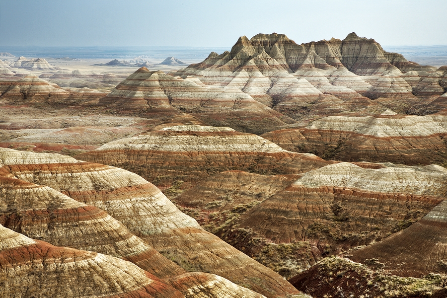
When I was a seven year-old kid, my parents took me on an extended, wild road trip adventure across the North American continent, from the east coast to the west coast. One of our adventure breaks along the way was to roam about the Badlands of North Dakota. It was a dusty, barren place, but my mother managed to unearth a treasure amid the empty landscape. She saw something sticking up out of the hard-packed clay ground and kicked at it until it broke loose a little, and then she dug it up. It was a fossilized skull of some unidentified animal, complete with darkened molars and empty fang sockets. To this day, I still have no idea what this petrified animal could have been but it is small, fitting into the palm of my hand. This single event shaped my entire life's passion. When asked in grade 3 what I most wanted to be when I grew up, I stated confidently, "An archaeologist."

I did not become an archaeologist, but an artist specializing in fibre and material studies, predominantly utilizing bone, flesh, hair and fur in my practice. Till now, I have worked with dead matter, bringing it back to life through mythic, artistic reconstruction. This is both a spiritual and physical practice. Naturally my next step is to literally bring materials to life through wet biology processes, to construct new semi-living (and perhaps still mythic) artistic objects. It amazes me that I have the tools and resources to do this as an artist-researcher.

My research will be focused at Concordia University this academic year, partially independently in my private studio, as well as through peer discussion, hopefully some consultation with professor Tagny Duff, as well as eventually (maybe next year) in the new tissue engineering lab that Tagny is establishing at Concordia if she will agree to have me in!

I'm particularly interested in this as a calcium (bony) growth that resembles knitting. This is a fossilized heliastar (starfish).
In the next post, I will share my actual studio and lab work around all of this so far.